Label-free imaging: What is it good for? Absolutely most things!
Craig Goergen
Say it again! And while one of the plenary talks in the Opening General Session given by Sunny Xie (Monday, April 15th, 8:00 to 9:45am in the Ali’Ii Ballroom) titled "Label-Free Vibrational Imaging for Medicine" might be a little less funky, I am sure it will be equally as entertaining.
Dr. Xie is the Mallinckrodt Professor of Chemistry and Chemical Biology at Harvard University and is considered by many to be a founding father of the field of single-molecule enzymology. His group has also made significant contributions to the development of Coherent Anti-Stokes Raman Scattering (CARS - a dye-free method in which image structures are characterized by intrinsic vibrational contrast of their molecules). The advantage of this method is that it does not require labeling and the sample remains mostly unaffected.
Dr. Xie's plenary talk in Hawaii will focus on a similar technique referred to as stimulated Raman scattering (SRS) microscopy. In spontaneous Raman scattering, only one laser beam illuminates the sample. This laser can create a signal that is generated at both the Stokes and anti-Stokes frequencies due to energy transfer between the photons and the molecules during their interaction. In SRS, however, two laser beams coincide on a sample. When the difference in frequency between the beams matches a particular molecular vibrational frequency, the Raman signal can be amplified by this stimulated excitation. SRS is also a label-free and noninvasive imaging technique that uses vibrational spectroscopy as the contrast. In other words, SRS measures vibrational energy levels that are associated with the specific chemical bonds in a sample (see Figure 1). Like a fingerprint, the sample's spectrum is unique, meaning that vibrational spectroscopy can be used for identification, characterization, and structure elucidation. SRS does not produce a background signal since signal cannot be produced when the laser frequency difference does not match the absorbed vibrational resonance.
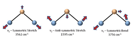
Figure 1: The independent vibrational modes of a water molecule (H20). Each of these has a corresponding fundamental resonant frequency. Image reproduced from: http://www.hsimagazine.com/article.php?article_id=248.
Several recent advancements in SRS have significantly improved the technique's sensitivity, selectivity, robustness, and overall cost. This is opening a wide range of biomedical applications, including the study of lipid metabolism, drug delivery, and tumor diagnosis. Below are images that highlight a relatively new contrast mechanism for imaging tumors. By making use of differences in lipid and protein density, cell nuclei morphology can be derived. This provides contrast analogous to H&E histology, making tumor margins easily visible. Thus, clinicians may one day soon be able to analyze images with subcellular spatial resolution in unstrained fresh tissue taken directly from the operating room.
Figure 2 highlights SRS images of both primary and metastatic tumors, displaying both the green lipids and blue nuclei. Not only can the tumor margin be clearly identified, but the margins of both primary and metastatic tumors appear different. Comparison between H&E stains and SRS images clearly establishes the chemical selectivity of this technique. Further efforts to establish the use of this technique for virtual histology during brain tumor surgery are underway.
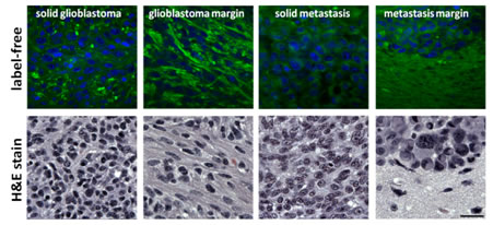
Figure 2: SRS images of glioblastoma multiform and breast cancer brain metastasis highlight tumor margins. Image reproduced from: http://bernstein.harvard.edu/research/carsapps.html.
Raman microscopy can also be used to study the dynamics of drug penetration through the skin. Figure 3 shows the time-lapse SRS images of drug penetration. The decrease from the skin surface is comparable with data from more invasive and labor-intensive tape-stripping results.
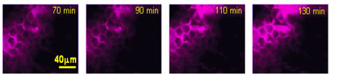
Figure 3: Imaging the penetration of ketoprofen across the stratum corneum layer of the skin using SRS contrast at 70, 90, 110, and 130 minutes after application. Image reproduced from: http://bernstein.harvard.edu/research/carsapps.html.
I am looking forward to Dr. Xie's plenary talk in Hawaii (which again will be on Monday, April 15th, 8:00 to 9:45am in the Ali’Ii Ballroom). I'll be there - hope you will as well!
