About
27 August 2019
Computational Approach Speeds Up Advanced Microscopy Imaging
Faster imaging means scientists can observe hundreds of neurons at once with two-photon microscopy
WASHINGTON — Researchers have developed a way to enhance the imaging speed of two-photon microscopy up to five times without compromising resolution. This record-fast imaging speed will allow scientists to observe biological phenomena that were previously too fleeting to image with current state-of-the-art advanced microscopy.
Caption: Researchers have developed a system that increases the imaging speed of two-photon microscopy up five times without compromising resolution. On the left, is a CAD model of the compact custom built two-photon microscopy system. The free-space optics inside is pictured on the right.
Credit: Shih-Chi Chen from The Chinese University of Hong Kong
In The Optical Society (OSA) journal Optics Letters, researchers led by Shih-Chi Chen from The Chinese University of Hong Kong describe how they combined a computational imaging approach known as compressive imaging with a faster scanning method. They used the new method to acquire two-photon microscopy images of a pollen grain in less than one second. This would take five times as long using the traditional approach.
“This new compressive sensing-based two-photon microscopy method will be useful for visualizing a neural network or monitoring activity from hundreds of neurons simultaneously,” said Chenyang Wen, first author of the paper. “Typically, neurons transmit signals on a time scale of 10 milliseconds, which conventional systems are too slow to follow.”
Speedier scanning
Two-photon microscopy works by delivering ultrafast pulses of infrared laser light to the sample where it interacts with tissue or fluorescent labels that emit signals used to create an image. It is extensively used for biology research because of its ability to produce high-resolution, 3D images up to a depth of one millimeter. These advantages, however, come with a limited imaging speed because the low-light conditions call for point detectors that require point-by-point image acquisition and reconstruction.
To speed up imaging, the researchers previously developed a multi-focus laser illumination method that uses a digital micromirror device (DMD), a type of low-cost light scanner typically used in projectors. “It was thought that these DMDs could not work with ultrafast lasers,” said Chen. “However, we recently addressed this issue, which has enabled application of DMDs in ultrafast laser applications that include beam shaping, pulse shaping, fast scanning and two-photon imaging.”
The DMD generates five to 30 points of focused laser light on randomly selected locations within a specimen. The position and intensity of each point of light are controlled by a binary hologram that is projected onto the device. During each measurement, the DMD reflashes the hologram to change the position of each focus and records the intensity of the two-photon fluorescence with a single-pixel detector. Although, in many ways, DMD multi-focus scanning is more flexible and faster than traditional raster scanning, the speed is still limited by the rate at which the device can form light patterns.
Combining methods brings faster imaging
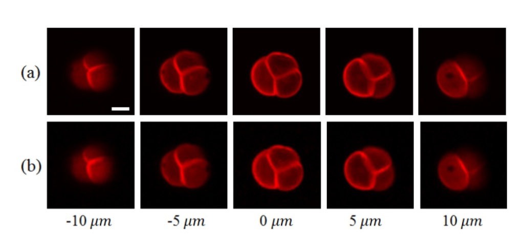
Caption: The researcher compared two-photon microscopy images of a pollen grain using traditional raster scanning (a) and their new compressive imaging approach (b). The raster-scanning imaging time was 2.2 seconds while the compressive imaging time required only 0.55 seconds.
Credit: Shih-Chi Chen from The Chinese University of Hong Kong
In the new work, the researchers further increase the imaging speed by combining multi-focus scanning with compressive sensing. This computational approach enables image reconstruction with fewer exposures because it carries out sampling and image compression in a single step and then uses an algorithm to fill in the missing information. For two-photon microscopy, it allows a specimen to be reconstructed using 70 to 90 percent fewer exposures than traditional approaches.
After conducting a simulation experiment to demonstrate the new method’s performance and to identify optimal parameters, the researchers tested it with two-photon imaging experiments. These experiments demonstrated the technique’s ability to produce high-quality 3D images with high imaging speeds from any field of view. For example, they were able to acquire images from five layers in a pollen grain, with each layer measuring 100 × 100 pixels, in just .55 seconds. The same images acquired with raster scanning took 2.2 seconds.
“We achieved a 3 to 5 times enhancement in imaging speed without sacrificing the resolution when imaging arbitrarily selected regions in 3D specimens,” said Wen. “We believe this new compressive sensing-based approach will be useful to use with approaches such as optogenetics in which light is used to control neurons and will lead to new discoveries in biology and medicine.”
The researchers are working to further improve the speed of the reconstruction algorithm and image quality. They also plan to use the DMD platform with other advanced imaging techniques such as wave front correction, which allows deep tissue imaging.
Paper: C. Wen, M. Ren, F. Feng, W. Chen, S.-C. Chen, “Compressive sensing for fast 3-D and random-access two-photon microscopy,” Opt. Lett., 44, 17, 4343-4346 (2019).
DOI: https://doi.org/10.1364/OL.44.004343.
About The Optical Society
The Optical Society (OSA) is dedicated to promoting the generation, application, archiving, and dissemination of knowledge in optics and photonics worldwide. Founded in 1916, it is the leading organization for scientists, engineers, business professionals, students, and others interested in the science of light. OSA’s renowned publications, meetings, online resources, and in-person activities fuel discoveries, shape real-life applications and accelerate scientific, technical, and educational achievement.
About Optics Letters
Optics Letters has been publishing high-impact research in the field of photonics for over 45 years and offers rapid dissemination of new results in all areas of optical science with short, original, peer-reviewed communications. Optics Letters accepts papers that are noteworthy to a substantial part of the optics community. Published by Optica Publishing Group and led by Editor-in-Chief Miguel Alonso, Institut Fresnel, École Centrale de Marseille and Aix-Marseille Université, France, University of Rochester, USA. For more information, visit Optics Letters.
Media Contact
