About
20 July 2016
New Probe Developed for Improved High Resolution Measurement of Brain Temperature
20 July 2016
New Probe Developed for Improved High Resolution Measurement of Brain Temperature
Improved accuracy could allow researchers to measure brain temperature in times of trauma when small deviations in temperature can lead to additional brain injury
WASHINGTON — The brain is the most temperature-sensitive organ in the body. Even small deviations in brain temperature are capable of producing profound effects—including behavioral changes, cell toxicity, and neuronal cell death. The problem faced by researchers and clinicians is how to measure and understand these changes in the brain and how they are influenced by complex biochemical and physiological pathways that may be altered by disease, brain injury or drug abuse.
In a new paper published in Biomedical Optics Express, from The Optical Society (OSA), Stefan Musolino of the University of Adelaide and the ARC Centre of Excellence for Nanoscale BioPhotonics, Australia, and his colleagues describe a new optical fiber-based probe capable of making pinpoint brain temperature measurements in moving lab animals.
 |
||
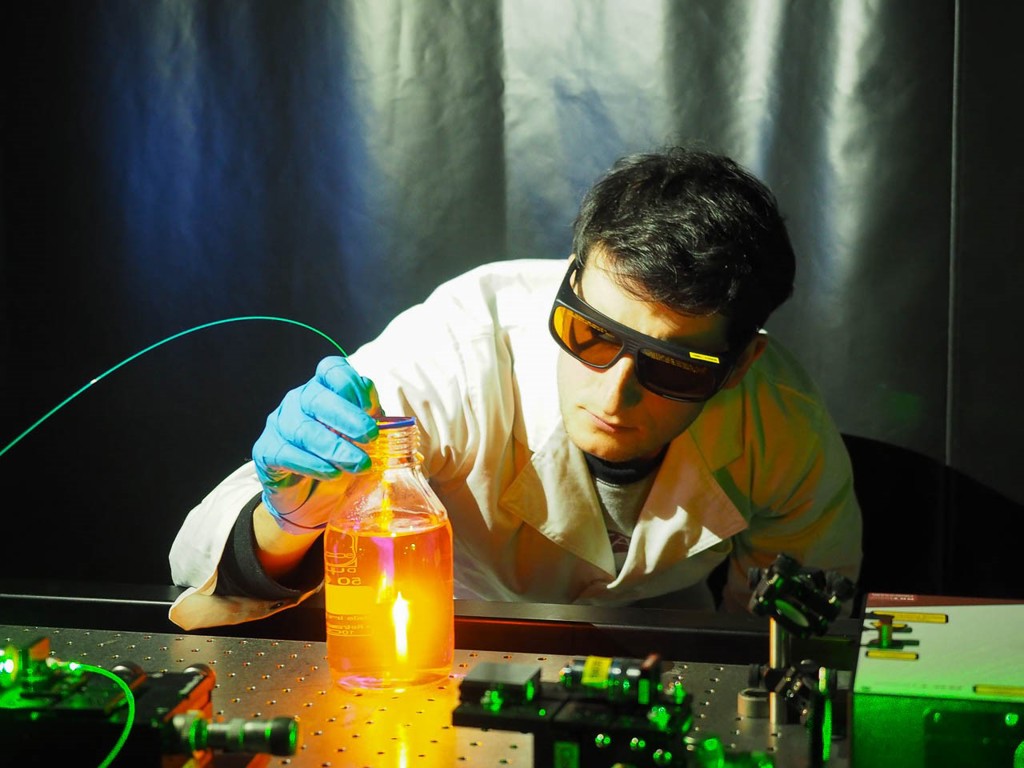
| The optical fiber probe can generate and harness light to detect temperature changes in biological systems. Image Credit: Erik Schartner & Stefan Musolino University of Adelaide |
“Within our center we house physicists, chemists, and medical researchers and one of the interests of our center’s ‘Origin of Sensation’ theme is temperature change in the central nervous system,” Musolino said. “It is only recently that more studies in my area of research— drug-induced hyperthermia— have started looking at changes in brain temperature in addition to changes in core body temperature within drug-treated animals. We wanted to further investigate these drug-induced brain temperature changes using center developed probes in order to develop a better understanding of the mechanisms driving them.”
The probe developed by Musolino and his colleagues consists of an optical fiber, sheathed within a protective sleeve and encased within a 4-millimeter-long 25-gauge needle. The end-face of the approximately 2-mm-long probe tip is dipped into molten glass made of tellurite, doped with a small amount of the rare-earth oxide erbium. When inserted into the brain, the color of the light emitted from the erbium ions will vary depending on the temperature of the surrounding tissue; the temperature of that tissue can thus be determined by monitoring the light of these color changes. This method allows for measurements to be performed with a precision of a fraction of a degree (0.1°C).
“The area that can measure temperature is less than 125 micrometers in size,” said study co-author Erik Schartner “making it highly spatially precise and able to isolate temperature readings from very small brain areas.” The researchers say it is possible to make the temperature-sensing area of the probe tip smaller still — as small as a few microns across — by modifying the probe’s design.
The probe’s immediate application will be to investigate changes in brain temperature within moving lab animals exposed to certain drugs of abuse, such MDMA (or ‘ecstasy’). “We will also look at the possible therapeutic properties of the tetracycline antibiotic minocycline and its ability to attenuate the changes in temperature caused by the administration of MDMA,” said Musolino. “In the future we will also be looking into combining this probe with other optical sensors in the hopes of developing new optical fiber-based sensing techniques for use in medical science labs that are examining real-word medical problems.”
Eventually, a fully developed probe could be used in human brain temperature monitoring after traumatic brain injury, stroke or hemorrhage — times when the brain is extremely sensitive and small deviations in temperature can lead to additional brain injury.
“Continuous monitoring of brain temperature after brain injury would allow for the effects of hyperthermia management techniques such as anti-pyretics — drugs that reduces fever — and hypothermia to be observed and evaluated by clinicians in real time,” Musolino said. “These new tools and this deeper understanding will ultimately give us better understanding of the brain and how to more quickly react to brain injury.”
Paper: Stefan Musolino, Erik P. Schartner, Georgios Tsiminis, Abdallah Salem, Tanya M. Monro, and Mark R. Hutchinson, "Portable optical fiber probe for in vivo brain temperature measurements," Biomed. Opt. Express 7, 3069-3077 (2016). DOI: 10.1364/BOE.7.003069.
About Biomedical Optics Express
Biomedical Optics Express is OSA’s principal outlet for serving the biomedical optics community with rapid, open-access, peer-reviewed papers related to optics, photonics and imaging in the life sciences. The journal scope encompasses theoretical modeling and simulations, technology development, and biomedical studies and clinical applications. It is published by The Optical Society and edited by Christoph Hitzenberger of The Medical University of Vienna. Biomedical Optics Express is an open-access journal and is available at no cost to readers online at: OSA Publishing.
About The Optical Society
Founded in 1916, The Optical Society (OSA) is the leading professional organization for scientists, engineers, students and entrepreneurs who fuel discoveries, shape real-life applications and accelerate achievements in the science of light. Through world-renowned publications, meetings and membership initiatives, OSA provides quality research, inspired interactions and dedicated resources for its extensive global network of optics and photonics experts. For more information, visit osa.org/100.
Media Contacts:
mediarelations@osa.org
