Day 2 of the Future Microscopy Incubator
Ivo Leite, University of Dundee, UK
Day Two of the Future Microscopy Incubator started with coffee and biscuits. After settling in, the participants were divided into two groups for breakout discussions. For two hours, each group discussed the challenges and strategic aims to make future techniques in microscopy either “Faster & Smaller” or “Broader & Deeper.” By the end of the morning, the participants reconvened to present their conclusions from each breakout session.
Romain Laine (Univ. Cambridge, UK) and Michelle Peckham (Univ. Leeds, UK) summarised the conclusions from the breakout discussion addressing the technical and practical limits of spatial and temporal resolution. One of the main questions was: is there such a thing as fast enough or small enough? The answer depends on the particular application and scale (spatial and temporal) of the phenomena under analysis. Furthermore, it is important to note that the two can be interchangeable, according to the technique used. Single-molecule localisation microscopy transfers spatial to temporal resolution, while streak cameras do the opposite. Several strategies have been used to increase imaging speed. In high-speed volumetric imaging, multiplane imaging can bring improvements. Another limiting factor in fluorescence microscopy techniques can result from the labelling. Dyes requiring lower photon budgets could push the temporal resolution towards smaller values, and there is a hope that quantum-dot engineering could bring even better results in the near future. Other approaches to faster imaging can be image de-noising to increase the signal-to-noise ratio (SNR), or building prior information in the data analysis to get away with lower SNR. Regarding the development of technology, achieving modularity in hardware and in hardware control by agreeing on standards and common requirements – not only among the research community but also involving manufacturers – could speed up the development of new microscopes. Following the example of OpenSPIM, the adaptive optics community could greatly benefit from the synergies offered by such an open access platform.
The conclusions of the other breakout discussion were summarised by Tomas Cizmar (Univ. Dundee, UK) and Ioannis Papadopoulos (Charité and Humboldt Univ., Germany). The trade-offs between spatial resolution vs imaging depth, as well as in field-of-view vs volume of data and cost of components dominated most of the discussion. The influence of complex photonics in future microscopes was also addressed. The theoretical limits for the imaging depth in tissue samples is estimated to be around 2 mm in the ballistic regime, but the combination of photo-acoustics with wavefront shaping in the diffusive regime could take this theoretical limit up to 2 cm. It could, however, take decades to reach half of this value, noted Allard Mosk (Utrecht Univ., Netherlands), due to the complexity of such techniques. On the other hand, in the opinion of Jacopo Bertolotti (Univ. Exeter, UK) there is still a lot of new physics to be learned in this particular field, so it might happen that these limits will be pushed even further.
In the afternoon, Gail McConnell (Univ. Strathclyde, UK) synthesised the main questions which arose over the two-day meeting. A need for multi-colour super-resolution microscopy was identified and may constitute a research opportunity. The different methods currently used to quantify resolution in super-resolution microscopy – maximum likelihood, least-squares fitting, and centroid – all give different outcomes, and agreeing on standardisation is paramount. Devising new ways of using prior information can significantly reduce SNR and improve resolution, and here perhaps the community could learn from high-energy and particle physics which have a strong accumulated experience in statistical methods of data analysis. Among the recommendations for future practice in the field, the more routine use of bead samples for checking commercial microscope performance should be adopted, although care must be taken in the choice of bead. It also seems beneficial to further discuss – and here involve the funding agencies in the dialogue – the establishment of standard guidelines and requirements (e.g. optical power at the specimen and using a standard specimen for photobleaching) in the dissemination of results. Finally, a list of recommendations was compiled for the interfacing software between the microscopes and the end users. These include the storing of metadata in an open format (which ideally should include all information on the microscope, the sample, and imaging parameters), store all image processing, manipulation and analysis results, and help to inform and educate the user.
To conclude the meeting, Mat Wasley from the Knowledge Transfer Network (UK) gave an overview of the present and upcoming funding landscape in Europe and in the UK, with particular emphasis on the opportunities specifically targeting the photonics-related disciplines. The uncertainty scenario surrounding Brexit couldn’t be left out, with its future impact being at present difficult to grasp. The current policies of the European and British funding agencies seem to continue to lean towards applied research, including in particular those bringing potential impact in the fields of health and life Sciences. This seems to mean that there are – and there will continue to be in the next few years – plenty of opportunities to support research in the field of microscopy.
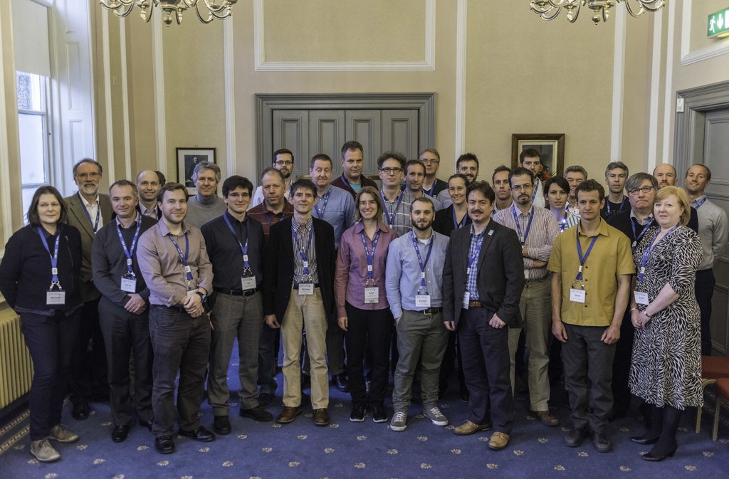
A sense of accomplishment was noticeable in the room as the hosts closed the meeting. The participants were visibly pleased with the fruitful – and intensive – discussions offered by the unusual format that is characteristic of an OSA Incubator Meeting, and it was the first such meeting for the vast majority of them. Overall a great success for this last scientific meeting of the year in which The Optical Society celebrates its first centennial.