How to fit a laser-scanning microscope into a 2mm diameter tube
Kyle Quinn
Optical microscopy can provide high-resolution images of cellular morphology and matrix organization, which can be utilized to diagnose disease or trauma. However, achieving an adequate signal-to-noise ratio at imaging depths exceeding 1mm is very challenging. As a result, the initial clinical applications for optical microscopy techniques have largely focused on skin pathology. One approach to unlocking a wider spectrum of clinical applications for biomedical optics is miniaturizing the distal end of microscopes into endoscopic probes.
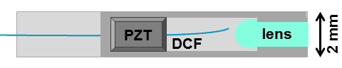
Over the last 20 years, researchers have been developing endomicroscopes to enable confocal or multi-photon microscopy in hard to reach places like the gastro-intestinal (GI) track. Dr. Xingde Li and his team of researchers in the Laboratory of Biophotonics Imaging Technologies at Johns Hopkins University have been leading the way by developing novel fiber-optic endomicroscopes to perform non-destructive, real-time optical biopsies. There are a number of engineering challenges associated with miniaturizing an objective lens and laser scanning system while still obtaining high-quality multi-photon microscopy images. Rapid scanning times, efficient light collection, minimal chromatic aberrations, and maintaining ultrashort pulses at the distal end of the fiber represent a few of the criteria that should be met when designing flexible endomicroscopes for multi-photon use. Dr. Li’s group has not only met these challenges, but packaged their probe within a slim 2mm-diameter hypodermic tube. They utilize a double-clad fiber (DCF) for both transmitting excitation and emission light and a small miniaturized compound lens at the distal end of their probe. Rapid scanning in a 2D spiral pattern is achieved by actuating the optical fiber cantilever with a piezoelectric (PZT) resonator.
In his plenary lecture at BIOMED 2014, Dr. Xingde Li will be discussing the continued development of this technology and some of its potential applications. His group is currently assessing the diagnostic value of this technology for quantifying the risk of pre-term birth (PTB)- a major cause of infant mortality. Because structural and biochemical changes within the cervix may precede uterine contractility, their endomicroscope may provide quantitative biomarkers to identify woman at risk for PTB. Using a mouse model, their initial work has focused on SHG imaging of collagen organization using their probe. At BIOMED, Dr. Li will discuss more recent efforts in this mouse model to acquire additional information through the collection of two photon excited fluorescence from NADH, FAD, and elastin.
The quantification of these endogenous biomarkers through endomicroscopy has applications beyond cervical assessments, including the potential for non-invasive optical biopsies of the small intestine, colon, kidney, and pancreas. Although much effort has been placed on translating endomicroscopy technology to the clinic, Dr. Li also emphasizes that these devices can also be an enabling technology in basic science research. Research involving 3D in vitro tissue cultures or in vivo animal models can benefit from non-destructive, depth-resolved biochemical and microstructural assessments. His endomicroscope may be particularly amenable to researching infectious diseases, in which the distal end of the technology could reside within an enclosed space designed for higher biosafety levels. The neuroscience community has also been an early adopter of multi-photon microscopy technology, and Dr. Li will also touch on potential applications in brain imaging on awake animals. Whether you are interested in microscopy applications for the laboratory or the clinic, be sure to check out Dr. Li’s plenary session in April.
