About
19 September 2016
Promising New Imaging Tool Allows Surgeons to Detect Malignant Tissue During Breast-Conserving Surgery for Breast Cancer
19 September 2016
Promising New Imaging Tool Allows Surgeons to Detect Malignant Tissue During
Breast-Conserving Surgery for Breast Cancer
Combining wide-field micro-elastography with Optical Coherence Tomography (OCT), an Australian research team have developed a new imaging tool to detect malignant tissue during surgery that offers the potential to dramatically reduce the number of reoperations for patients with breast cancer
WASHINGTON — Every year, an estimated 1.6 million women are diagnosed with breast cancer worldwide. It is one of the most common forms of cancer to affect women, second only to skin cancer. It is also deadly, killing an estimated 522,000 women annually.
The development of mammography technologies has aided in the detection of earlier stage breast cancers. As a result, it is often not necessary to remove the entire breast, and there has been an increasing trend towards wide-local excisions, a type of breast-conserving surgery that involves the removal of the lump or tumor from the breast. During these operations, surgeons aim to remove the tumor, along with a thin rim of healthy tissue, known as the “surgical margin,” to ensure that the tumor does not reoccur. To be certain that all the malignant tissue has been removed, samples are taken at the margin for pathology testing.
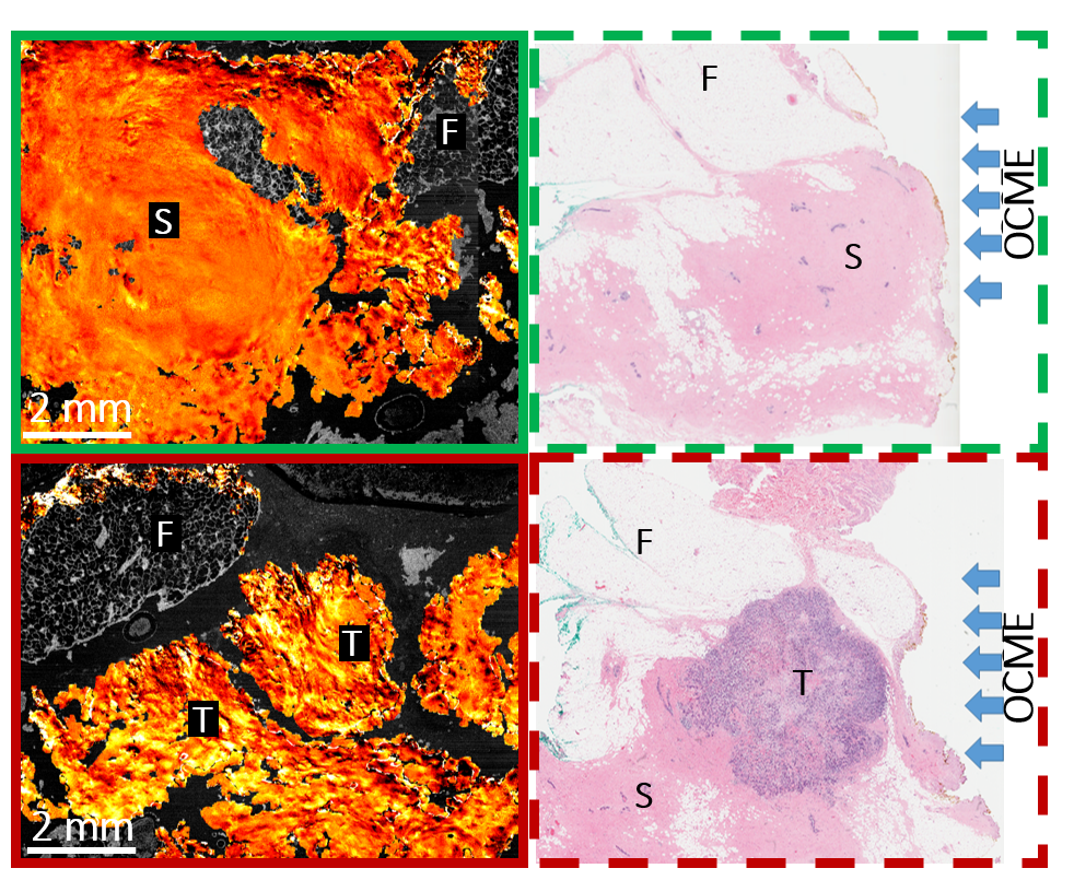 |
| 'Magnified images from wide-field OCME of freshly excised human breast tissue. Green solid square: uniform strain texture in OCME image indicative to healthy straoma (S). Red solid square: heterogeneous strain texture in OCME image indicative of malignant tumour (T). Dashed squares show corresponding histology. Both regions contain regions of fat tissue (F).' Image Credit: Wes Allen |
“The challenge with the results of these tests is that they are often only available days after the surgery,” said Wes Allen, a researcher and electronic engineering doctoral student at the University of Western Australia. “If malignant tissue is discovered, the patient must undergo surgery again to remove it. It’s estimated that 20 to 30 percent of breast-conserving surgery patients must undergo a second surgery.”
“These second surgeries are a large financial burden for healthcare systems,” Allen said. “They also force patients to endure the emotional toll of undergoing surgery again and can delay other related treatments.” Working closely with Brendan Kennedy and David Sampson, professors of electronic engineering and Christobel Saunders, a professor of surgery at the University of Western Australia, Allen has developed a new tool that allows surgeons to detect malignant tissue during surgery. The researchers describe their technique, which they have termed “Optical coherence micro-elastography,” (OCME), in a paper published this week in the journal Biomedical Optics Express, from The Optical Society (OSA).
“This tool will provide surgeons with feedback about whether the margin has malignant tissue while the patient is still in the operating room,” Allen said.
The new tool builds on a pre-existing medical imaging system called optical coherence tomography (OCT) which generates 3D, high-resolution images based on how different portions of a tissue sample reflect laser light. OCME utilizes the OCT imaging system to measure how different portions of tissue respond to being physically compressed. The amount of compression within the tissue is related to its mechanical properties. OCME then overlays the mechanical properties onto the OCT image. The hybrid image that is generated allows surgeons to better differentiate between malignant and healthy tissue.
The research group, whose work is funded by Australia’s National Health and Medical Research Council, Australia's National Breast Cancer Foundation, and the Australian Research Council, had previously proven the validity of this new technique with a small actuator, the device that briefly compresses the samples. With their current work, Allen and his collaborators integrated a larger, wide-field actuator into the set-up and developed a new protocol to speed up the scanning process.
“By moving to a wide-field actuator, we can scan the entire face of specimens excised during wide-local excision,” Allen said. “This means that we have a bench top system that can have clinical relevance.”
Ultimately, Allen and his fellow researchers believe that their technology could be translated into a handheld probe that surgeons can use to directly inspect margins within the patient for malignant tissue.
While the ingenuity of this new tool has the potential to spare thousands of breast cancer patients the burden of a second surgery, Allen is quick to credit the collaborative, cross-disciplinary ethos of the research group for their success to date.
Allen continues, “By working closely with surgeons and pathologists, we develop a good understanding of what they need. As engineers, we can develop fantastic tools with high resolution, but if they’re not solving the clinical problem that the surgeons tell us about, they’re not going to make an impact.”
Paper: Wes M. Allen, Lixin Chin, Philip Wijesinghe, Rodney W. Kirk, Bruce Latham, David D. Sampson, Christobel M. Saunders, and Brendan F. Kennedy, "Wide-field optical coherence micro-elastography for intraoperative assessment of human breast cancer margins," Biomed. Opt. Express 7, 4139-4153 (2016).
DOI: 10.1364/BOE.7.004139.
About Biomedical Optics Express
Biomedical Optics Express is OSA’s principal outlet for serving the biomedical optics community with rapid, open-access, peer-reviewed papers related to optics, photonics and imaging in the life sciences. The journal scope encompasses theoretical modeling and simulations, technology development, and biomedical studies and clinical applications. It is published by The Optical Society and edited by Christoph Hitzenberger, Medical University of Vienna. Biomedical Optics Express is an open-access journal and is available at no cost to readers online at: OSA Publishing.
About The Optical Society
Founded in 1916, The Optical Society (OSA) is the leading professional organization for scientists, engineers, students and entrepreneurs who fuel discoveries, shape real-life applications and accelerate achievements in the science of light. Through world-renowned publications, meetings and membership initiatives, OSA provides quality research, inspired interactions and dedicated resources for its extensive global network of optics and photonics experts. For more information, visit osa.org/100.
Media Contacts:
mediarelations@osa.org
