About
13 December 2012
No More Lying About Your Age: Scientists Can Now Gauge Skin’s True Age with New Laser Technique
FOR IMMEDIATE RELEASE
Contact:
Lyndsay Meyer
The Optical Society
+1.202.416.1435
lmeyer@osa.org
No More Lying About Your Age: Scientists Can Now Gauge Skin’s True Age with New Laser Technique
New skin age index could provide test-bed for measuring effectiveness of ‘anti-aging’ skin products
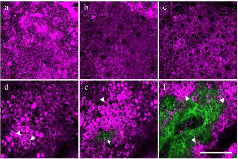
Figure 1: This series of harmonic generation microscopy images shows the skin cells of a 24-year-old subject at increasing depths, ranging from the outermost layer of skin (a) to approximately 300 millionths of a meter deep (f). The magenta areas, generated from third harmonics, show skin cells and their nuclei. The green areas, generated from second harmonics, show fibers made of the protein collagen. Credit: Biomedical Optics Express.
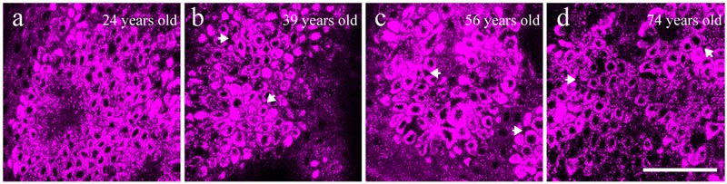
Figure 2: This shows images of basal keratinocytes, the most common cells in the outermost layer of skin. The images were taken of the forearms of a 24-year-old (a), a 47-year-old (b), a 59-year-old (c) and a 69-year-old (d). Compared to the skin cells of the youngest volunteers, the skin cells in older subjects were larger, more irregular in shape, and showed spaces between cells, as indicated by the white arrows in images b-d. Credit: Biomedical Optics Express.
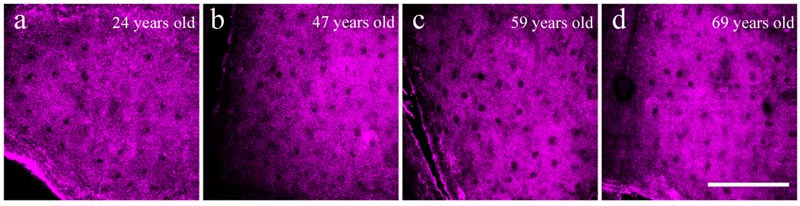
Figure 3: This shows images of granular skin cells, a type of cell found near the top of the outermost layer of skin, for a 24-year-old (a), a 47-year-old (b), a 59-year-old (c) and a 69-year-old (d). In contrast to the irregularities that developed in the basal cells of older subjects, the images of granular cells revealed no significant correlation between age and size or shape of the cells. Credit: Biomedical Optics Express.
WASHINGTON, Dec. 13, 2012—Wrinkles, dryness, and a translucent and fragile appearance are hallmarks of old skin, caused by the natural aging of skin cells. But while most of us can recognize the signs of lost youth when we peer into the mirror each morning, scientists do not have a standardized way to measure the extent of age damage in skin. Now a group of Taiwanese researchers has used a specialized microscope to peer harmlessly beneath the skin surface to measure natural age-related changes in the sizes of skin cells. The results, which are published in the Optical Society’s (OSA) open-access journal Biomedical Optics Express, can be used to study the general phenomenon of skin aging and may help provide an index for measuring the effectiveness of ‘anti-aging’ skin products.
In the study, Chi-Kuang Sun, a distinguished professor at National Taiwan University and chief director of the university’s Molecular Imaging Center, along with medical researcher and dermatologist Yi-Hua Liao and colleagues, evaluated 52 subjects ranging in age from 19 to 79 years old. The researchers focused a brief burst of infrared laser light into the skin of the subjects’ inner forearms, an area that is generally protected from sun damage, which accelerates natural aging. The beam penetrated to a depth of about 300 millionths of a meter, or approximately where the epidermis (the upper layer of skin) and the dermis (the lower layer) meet.
The researchers used a technique known as harmonic generation microscopy (HGM), which has previously been used to study developing embryos. In the procedure, a concentrated beam of photons is sent into a material. The photons naturally oscillate at a particular frequency, and as they interact with the material, they generate “harmonics” – vibrations that are multiples of the original frequency, which are characteristic of the material structure and properties. For example, the second harmonic is twice the original frequency and the third harmonic is three times the original frequency. In an imaging system, harmonics can reveal different structures at very high resolution. In their study, the team scanned for reflected second and third harmonic photons, and from those measurements, produced a high-resolution 3-D map of the tissue that revealed structures within the skin cells.
Natural aging, the scanning showed, caused a significant increase in the overall size of cells known as basal keratinocytes – the most common cells in the outermost layer of skin – as well as in the sizes of their nuclei. However, other types of skin cells, known as granular cells, did not show a similar pattern. Thus, says Sun, the relative changes in the two types of cells can serve as an index for scoring natural or “intrinsic” skin aging –the aging of skin caused by programmed developmental or genetic factors.
“No one has ever seen through a person’s skin to determine his or her age from their skin,” says Sun. “Our finding serves as a potential index for skin age.”
A skin age index would provide a standardized, quantitative scale that could be used rate the true “age” of skin, from young (less age-related damage) to old (more age-related damage). The scale could give doctors another tool to monitor the overall health of skin—by investigating whether the skin of certain individuals or populations ages faster or slower than average, tracking the aging of an individual’s skin over time, or testing how effective anti-aging treatments are at slowing the rate of skin aging.
Intrinsic, or chronological, aging is different from extrinsic aging, which is caused primarily by sun exposure. “There are a lot of extrinsic factors that can accelerate the aging process, such as smoking, ultraviolet light, and stress,” says Sun. The researchers found that the extent of extrinsic skin aging in their study subjects varied depending on occupation, personal habits, and skin type, but because the researchers looked at skin on the sun-protected inner forearm, their findings provide a measure of the primarily genetically-based intrinsic skin aging.
“This could provide an index for someone who cares about the health of their skin and might also provide a test-bed for measuring the effectiveness of ‘anti-aging’ skin products,” Sun says. “Of course,” he and Liao joke, “you could set an HGM scanner at the entrance to a bar, so you can know whether a person is over 21 years old and permitted for entry.”
Paper: “Determination of chronological aging parameters in epidermal keratinocytes by in vivo harmonic generation microscopy,” Biomedical Optics Express, Vol. 4, Issue 1, pp. 77-88 (2012).
EDITOR’S NOTE: High-resolution images are available to members of the media upon request. Contact Lyndsay Meyer, lmeyer@osa.org.
About Biomedical Optics Express
Biomedical Optics Express is OSA’s principal outlet for serving the biomedical optics community with rapid, open-access, peer-reviewed papers related to optics, photonics and imaging in the life sciences. The journal scope encompasses theoretical modeling and simulations, technology development, and biomedical studies and clinical applications. It is published by the Optical Society and edited by Joseph A. Izatt of Duke University. Biomedical Optics Express is an open-access journal and is available at no cost to readers online at www.OpticsInfoBase.org/BOE.
About OSA
Uniting more than 180,000 professionals from 175 countries, the Optical Society (OSA) brings together the global optics community through its programs and initiatives. Since 1916 OSA has worked to advance the common interests of the field, providing educational resources to the scientists, engineers and business leaders who work in the field by promoting the science of light and the advanced technologies made possible by optics and photonics. OSA publications, events, technical groups and programs foster optics knowledge and scientific collaboration among all those with an interest in optics and photonics. For more information, visit www.osa.org.
